
D.Frolov,R.Purlys*, S.Bagdonas, R.Rotomskis, M.Gavutis
Laser Research Centre, Vilnius University, Sauletekio 9, 2040 Vilnius
Lithuania
*Physics Faculty, Vilnius University, Sauletekio 9, 2040 Vilnius, Lithuania
![]()
|
D.Frolov,R.Purlys*, S.Bagdonas, R.Rotomskis, M.Gavutis
|
|
|
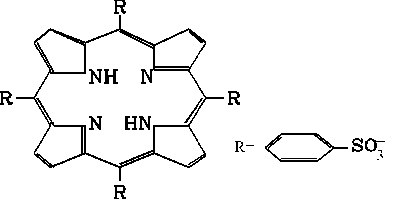
| Materials and Methods Meso-tetra(4-sulphonatophenyl)
porphine (TPPS4) was obtained from Porphyrin Products, Inc. (Utah, USA). This compound is
a porphyrin-type molecule possessing four phenyl substitutes at the meso positions with SO–3
groups located in para-position of phenyl ring (Fig.1). The
J-aggregates of TPPS4 were formed by dissolving in 0.1M HCl. .
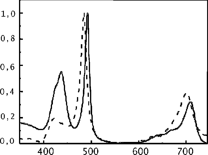
Absorption spectra of TPPS4 were measured by a S-1000 spectrometer (Ocean Optics Inc., USA). Sample for the X ray diffraction measurements was made by drying out a saturated TPPS4 solution in 0.1N HCl on an amorphous glass plate. Drops of J-aggregates solution were put on special plate and dried in air at room temperature. This procedure was repeated more than one hundred times to get the J-aggregates layer of about 0.3mm. During this procedure the J-aggregates had retained their spectral features unchanged (Fig.2).
Fig.2 Normalised absorption spectra of TPPS4 J-aggregates (solid line - in solution, dashed line - dried sample). The dependence of X ray scattering intensity on scattering angle was recorded by X-ray diffraction measuring system DPOH-2.0 . A X ray source with a copper anode was used for the irradiation of samples. (l Ka 1,2Cu=1,54Ĺ).
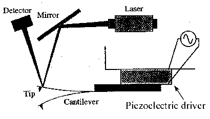
Atomic force microscopy measurements were made using Tapping mode Nanoscope III from
Digital Instruments. Image is forming by placing the tip in contact with the surface and then lifting it off the surface to avoid dragging across the surface (Fig.3). Tapping mode imaging is implemented in ambient air by oscillating the cantilever assembly at or near the cantilever’s resonant frequency using a piezoelectric crystal. The piezo motion causes the cantilever to oscillate with a high amplitude (typically greater than 20nm) when the tip is not in contact with the surface. The oscillating tip is then moved towards the surface until it begins to lightly touch, or “tap” the surface.
Fig.3 Principal scheme of Atomic force microscopy Tapping mode equipment
As the oscillating cantilever begins to intermittently contact with the surface, the amplitude of cantilever oscillation decreases because the surface limits cantilever’s motion. The reduction in oscillation amplitude is used to identify and measure surface features. Phase imaging is an extention of Tapping mode AFM. The resolution of phase imaging is compareble to the full resolution of Tapping mode AFM. Phase imaging can also be used as a real-time contrast enhancement technique. Because phase imaging highlights edges and is not affected by large-scale height differences, it provides clearer observations of fine features, such as grain edges, which can be obscured by rough topography. Sample for AFM measurements was made by drying one drop of saturated TPPS4 and HCl solution (pH1) on silicon vafer. This wafer had 10-20Ĺ oxide layer on top. |
![]()
![]()
|
|
![]()

