R.Rotomskis,
V.Vaicaitis, D.Frolov, S.Bagdonas,
Laser Research Centre, Vilnius University, Sauletekio 9, 2040 Vilnius
Lithuania
![]()
R.Rotomskis,
V.Vaicaitis, D.Frolov, S.Bagdonas, |
|
|
Results
|
Ground state absorption Emission Excited state absorption Fluorescencence kinetic spectroscopy |
||||||||||||||||||||||||||
| Ground state
absorption Figure 3 shows the ground electronic state absorption spectra of TPPS4 in aqueous, ethanol solutions and in aqueous solution in the presence of Triton-X100.
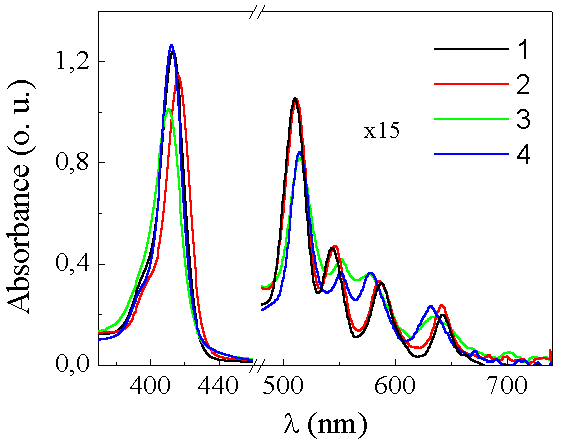
Fig.3 Absorption spectra of TPPS4 in: ethanol (black line, c=5*10-6M), aqueous solution with TR X100 (red line, pH=7.5, c=4*10-4M), aqueous solution at 4*10-4M and 5*10-6M (green and blue lines).
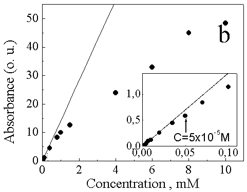
The absorption spectrum of TPPS4 in ethanol comprises the (p -p *) absorption bands characteristic of free-base etio type porphyrin. An intense near-UV Soret band (SB) consists of a peak at around 415 nm and a slight shoulder at higher energy due to B(0,0) and B(0,1) electronic transitions, respectively. Four weaker Q bands in the visible spectral region are located at 510, 544, 588 and 643 nm. .The absorption spectrum of TPPS4 in diluted aqueous solution at around neutral pH is typical for the most free bases monomeric porphyrins, similarly as that measured in ethanol solution. The absorbance of the SB and visible Q bands in aqueous solution at pH 7.5 shows linear dependence on the concentration of TPPS4 till 5*10-5 M (Fig.3a), indicating the dominance of free base monomers of TPPS4. Above this "thresholdĒ value the absorption spectrum of TPPS4 becomes different as it is displayed in Figure 4b. If a monomer (P(SO3-)4) - dimer (P2(SO3-)8) equilibrium present:
then the equation: (A0-A)=[(2e m-e d)(4KDC0+1-{1+8KDC0}1/2]/8KD should describe deviations from Beerís law (A0-A) in terms of dimerization konstant KD, the total TPPS4 concentration C0, and the molar extinction coefficients of the monomer ( e m) and dimer (e d). A0 is the calculated absorbance of the monomeric porphyrins and A is the observed absorbance of the porphyrin (monomer and dimer) at a given wavelength and concentration C0. The calculated dimerization constant of TPPS4 estimated from the absorption dependence on concentration (Fig.4) is 3x10³ M-1 and is close to the value presented in [19, 20].
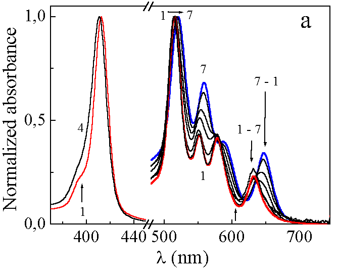
Fig.4 Concentration dependence of absorption of TPPS4 in aqueous solution at pH=7.5 and Bugerís law experiment. Compared with the spectra of monomeric porphyrin in ethanol solution (Fig.3) and monomerized porphyrin in aqueous solution in the presence of Triton-X100 (Fig.5), notable differences are observed in the spectrum of aqueous solution of TPPS4 at high concentration (Fig.4, curve 7): 1) B(0,0) band is weaker, broader and blue shifted by 3nm, 2) the B(0,0) B(0,1) peak intensities ratio is lower, and the B(1,0) Ė B(0,0) is slightly wider, 3) the Qx(0,0) Qx(1,0) and Qy(1,0) bands are shifted to the red, the Qx(1,0) - Qy(0,0) gap is smaller by 30% and all Q bands are broader for about 40%. These spectral changes might be explained by the formation of aggregates at high TPPS4 concentration in aqueous solution at pH 7.5.
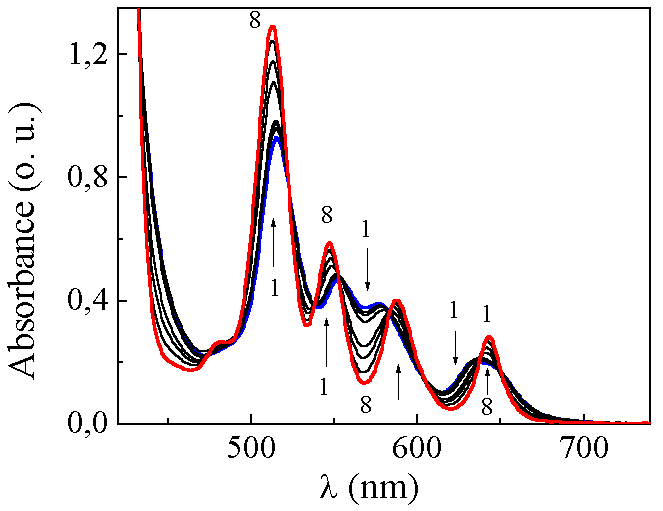
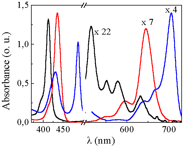
Fig.5 TR X100 concentration dependence (from 0% to 6%) of absorption of TPPS4 in aqueous solution. TPPS4 concentration of 1*10-3M, pH=7.5, absorption path length 1 cm. The structure of TPPS4 absorption spectra in aqueous solution strongly depends on pH (Fig.6). At around neutral pH, the absorption spectrum of TPPS4 consists of an intense SB at 413nm and four weak Q bands at 515,550, 578 and 631nm (Qy(1,0), Qy(0,0), Qx(1,0), Qx(0,0), respectively). In acidic solution (pH below 5,0) of low concentration the TPPS4 absorption spectrum in the visible spectral region changes to a three-band spectrum composed of an intense absorption band at 645 nm, and weaker bands at 597 and 550 nm. The SB is red-shifted to 435nm with respect to that at neutral pH (Fig.6). Since the existence of monoprotonated species of TPPS4 in acidic solutions is less expected [21, 22] these spectral changes might be attributed to the dication (H2+P(SO3-)4) (Fig.1c). Further decrease of solution pH results in an appearance of the new absorption bands at 490 and 706 nm. The SB undergoes a slight shift to the blue and a decrease in intensity like the rest of the absorption bands belonging to dication of TPPS4 (Fig.6). At pH 1.1 the TPPS4 absorption spectrum consists of the SB at 430 nm, two intense bands at 490 and 709 nm and three weak bands at 560, 640 and 670 nm (Fig.6). The band at 490 nm has a weak shoulder at 520 nm. The ratio between the intensities of the both absorption bands at 490 and 709nm and the absorption bands of dication varies depending on the total concentration of TPPS4 in acid solutions. The similar changes in absorption spectra can be induced by varying the ionic strength of acidic solution of TPPS4 and have been assigned to the formation of J-aggregates [23].
Fig.6 Absorption spectrum of TPPS4 in aqueous solution at different pH values. {pH=7.5 (solid), pH=3.5 (dash) and pH=1.1 (dot)}. Concentration 5*10-6M. Further decrease of solution pH to extreme values up to -1, results in a even reappearance of the absorption bands belonging to TPPS4 dication spectrum (Fig.7).
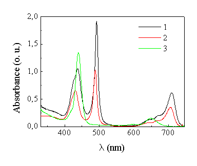
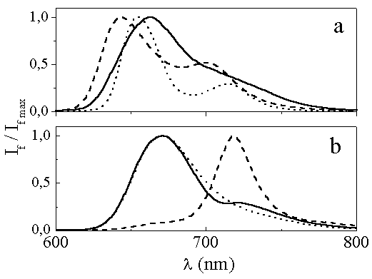
Fig.7 Absorption changes of TPPS4 depending on pH: curves 1,2 - pH 1.1 (in 0.1 N HCl), curve 3 - pH -1(in 40% H2SO4 aqueous solution). TPPS4 concentration: curve 1- ◊ 10-5 M, curves 2,3 - 5◊ 10-6 M, optical path length - 10 mm. The fluorescence spectra of TPPS4 in aqueous and ethanol solutions are presented in Fig.8a. In ethanol solution two bands with maxima at 655nm and 715nm corresponding to a (0Ė0) transition and its vibronic (0-1) transition can be seen in emission spectrum. Fluorescence bands of TPPS4 in aqueous solution are wider and blue-shifted by 9nm in comparison to those in ethanol solution. An increase in concentration of TPPS4 in aqueous solution caused the red shift of the main fluorescence maximum and disappearance of the second fluorescence maximum (Fig.8a). The aggregates formed at higher concentration in neutral solution show only one structureless emission band at 664nm due to the (0-0) transition, however the long tale on the red wing of the main fluorescence band might be assigned to the vibronic (0-1) transition. At pH 4 (Fig.8b) only one fluorescence band with a peak at 675nm due to the (0-0) transition is detected and attributed to the emission of the dication [24]. Some evidence of the coexistence of both dications and J-aggregates, which are formed at lower pH with increase in TPPS4 concentration or with presence of KCl to the aqueous solution, can also be drawn from the emission spectra in Fig.8b. Excitation at 420nm (where dications are the predominant absorbing fraction) gives rise to the emission of the dication whereas excitation at 488nm (an absorption band of J-aggregates) results in a different emission spectrum with a new distinctive peak at 720nm. This peak as the probably second weak peak, which is clearly seen at the same wavelength in the fluorescence spectrum of dication under excitation at 420nm, could be attributed to the fluorescence of J- aggregates.
Fig.8 Fluorescence spectrums of TPPS4 in: a) ethanol (concentration 10-6M) [dot] {l ex=420 nm} and aqueous solution at pH=7.5 (concentration 10-6M [dashed] and 10-3M [solid] {l ex=413 nm}; b) aqueous solution at pH=4 (c=5*10-6M) [dot] {l ex=420 nm} and aqueous solution at pH=2 (c=7.5*10-6M) {l ex=420 nm} [solid] ir {l ex=488 nm} [dashed].
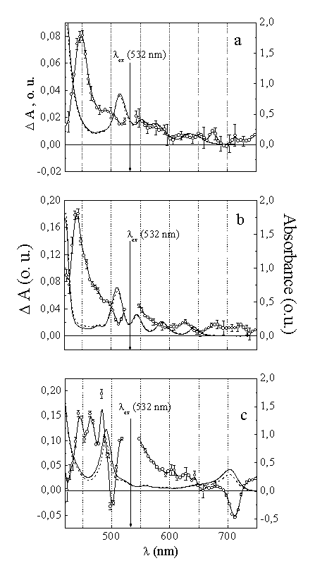
The absorption difference spectra of TPPS4 in aqueous solution at pH7.5 and in ethanol solution measured immediately after the excitation at 532nm exhibit bleaching of absorption bands with the maxima corresponding to the absorption bands of TPPS4 on the background of transient absorption. The transient spectrum (Fig.9a) recorded over a wide range of time, decayed monotonically in the aqueous solution of TPPS4, where equilibrium aggregates were detected. Such absorption changes are typical of the singlet excited state of porphyrins and were previously observed for heterogeneous hematoporphyrin [25]. The transient spectra presented in Fig.9a,b show that the singlet-excited state of TPPS4 in neutral aqueous solution and ethanol is characterized by a strong absorption in the 430-700 nm range, where the absorption cross-section from excited state is comparable or even greater than that of the ground state.The transient absorption spectrum recorded for TPPS4 in acidic solution (pH 3.5, in the presence of KCl) where the J-aggregates are formed. The instantaneous photobleaching of the J-aggregates bands at 490 nm and 708 nm is observed along with transient absorption on the blue and red side of the J-aggregates bands over the 430-750 nm domain.
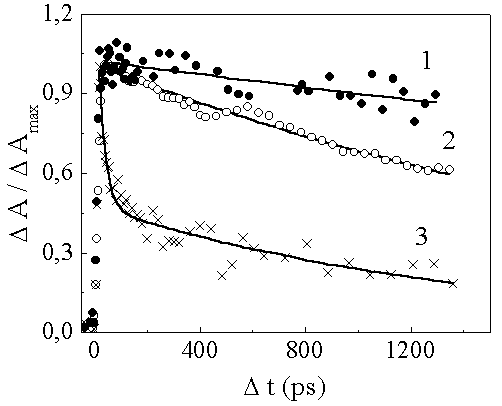
Fig.9 Absorption spectra of TPPS4 before (straight line) and after (dashed line) measurements as well as transient absorption difference spectrum measured with registration delay time D t=0 ps in: a) aqueous solution, pH=7.5, C=8x10⁻⁴M), b) ethanol, C=5x10⁻⁵M and c) aqueous solution, pH=3.5, C=4x10⁻⁴M. The absorption decay of TPPS4 J-aggregates induced by excitation can be fitted biexponentially with the time constants about t 1=27ps and t 2=1.44ns (Fig.10), or described by a three exponential decay pattern with the time constants t 1=24ps, t 2=290ps and t 3=2-3ns. In the case of the J-aggregates of TPPS4 the shortest time constant seems to be related to the exciton annihilation and a nanosecond time constant reflects the excitation energy relaxation when only one excitation is present in the J-aggregate.
Fig.10 Relaxation kinetics (excitation at l ex=532 nm, measured at l m=590 nm) of TPPS4 excited state in : ethanol (1), C=5◊ 10-5M (relaxation time t =7.9 ns), aqueous solution at pH=7.5 (2), C=8◊ 10-4M ,( t =3.1 ns) and aqueous solution at pH=3.5 (3), C=4◊ 10-4M, (t 1=27 ps and t 2=1.44 ns). Fluorescencence kinetic spectroscopyThe fluorescence of TPPS4 in aqueous solutions at different pH and concentrations was measured with the single photon counting fluorescence system described in the chapter Materials and methods. As it was shown earlier TPPS4 dissolved in the presence of H2SO4, where the value of pH is about -1.1 forms a homogenous solution of monomeric ionic species with characteristic absorption properties (Fig.7). The fluorescence decay kinetics measured in a wavelength range of 670-740 nm (excitaton at 398 nm) where fluorescence of protonated TPPS4 species was observed (Fig.8) shows a mono-exponential decay with a time constant of ca. 3.1 ns. In addition, the presence of a short component fluorescence decay was detected at around 660 nm. Its elucidation needs additional experiments to be performed. An increase of the pH from -1.1 to 1.19 results in the formation of J-aggregates as reflected by the appearance of the absorption band at 490 nm (Fig.6). At low concentrations (lower than 10-6 M) the dominant species in acidic solution is protonated monomer of TPPS4 which exhibits the monoexponential fluorescence decay with a time constant value close to that obtained at the pH-1.1 (Table 1).
Tab.1 Fluorescence decay constants obtained in TPPS4 aqueous solutions at different pH.
With the increase of TPPS4 concentration starting from 10-5 M the amount of J-aggregates in solution increases. In fluorescence decay kinetics it is reflected by the appearance of the short fluorescence decay component with the time constant about 0.27 ns in the spectral region of 690-740 nm (Tab.1), where steady state fluorescence spectrum attributed to J-aggregates was observed. Further increase of solution pH up to pH5 leads to the dissapearance of J-aggregates and the appearance of non-protonated species of TPPS4, which is reflected by coexistance of the absorption bands at 414 nm and 434 nm attributed to the non-protonated species (P(SO3-)4)(Fig.1a) and the protonated species (H2+P(SO3-)4)(Fig.1c). In fluorescence decay kinetics it is manifested by two fluorescence decay constants. In the spectral region where fluorescence of protonated species is dominant, the decay constant value is about 3.6 ns, which is the same as those obtained at the lower pH values. In the spectral region at 630-650 nm, where fluorescence of P(SO3-)4 is most intensive, decay constant is around 7.2s. The smaller decay constant value of P(SO3-)4 obtained in acidic medium in comparison with that in neutral solution might be caused by the fluorescence quenching effect induced by the protonated species present in the mixture. In neutral solutions at low TPPS4 concentration the value of the fluorescence decay constant is about 10ns. The presence of a short fluorescence decay component with a time constant around 0.7 ns might be explained by the formation of dimers with an increase of TPPS4 concentration (Fig.4). |
|||||||||||||||||||||||||||
![]()
![]()
|
|
![]()

