THE WHOLE HUMAN BODY DISTRIBUTION OF SOLAR ERYTHEMAL ULTRAVIOLET RADIATION
M.G. Kimlin‡†, A.V. Parisi†, J.C.F. Wong‡
†Centre for Astronomy and Atmospheric Research, Faculty of Sciences, University of Southern Queensland, TOOWOOMBA 4350 AUSTRALIA.
‡Centre for Medical and Health Physics, Queensland University of Technology, GPO Box 2434, BRISBANE 4001 AUSTRALIA.
Abstract
The paper presents a method for estimation of the erythemal ultraviolet (UV) exposure per unit area to four body zones, namely, the face, trunk, legs and arms employing only 23 polysulphone dosimeters. From these, the whole body exposure per unit area and the distribution of the body erythemal UV exposure were estimated. The overall picture of the body sites receiving high UV exposure was provided. On a day in winter at a sub-tropical site, the erythemal UV exposures per unit area over a ten hour period in a day ranged from 63 to 112 mJ cm-2 for the leg and arm zones respectively. Similarly, for a one hour period at approximately solar noon, the erythemal exposures per unit area ranged from 25 to 45 mJ cm-2. The exposures per unit area to the whole body were 34 and 90 mJ cm-2 for the one hour and ten hour exposures respectively.
Introduction
Human exposure to ultraviolet (UV) radiation has been associated in the formation of skin cancer (McCarthy and Shaw 1989, Longstreth et al 1995). UV exposure depends on many factors, such as ambient UV levels, activity undertaken, duration of activity, physical environment and UV protective measures. Human exposure to erythemal solar UV radiation has been previously investigated using polysulphone dosimeters (Diffey, 1989). These previous studies have investigated the erythemal UV exposure to specific human body sites using both manikins and human subjects. For example, Diffey et al (1977) investigated the UV to body sites under variable weather conditions during summer in England, while other studies (Gies et al 1988) have investigated the solar UV to body sites with season at a high latitude location in Australia. The erythemal exposure to specific body sites during various work and recreational activities has also been investigated by a number of authors, for example, Wong et al (1992), Gies et al (1995), Rosenthal et al (1991) and the level of UV exposure has been found to be correlated to human activity (Herlihy et al 1994). The effectiveness of hats in reducing the UV exposure to specific facial sites has also been investigated (Wong et al 1996, Diffey and Cheeseman 1992).
The exposures vary substantially from one body site to another site. For example, according to Diffey et al (1977) the range of exposures expressed as a percentage of the ambient varies from 30 to 73 for the cheek and lower sternum respectively. It would be uneconomical, inconvenient and difficult to assess the exposure to many body sites. The method presented in this paper allows estimation of the distribution of erythemal UV exposure to all body sites along with estimation of the whole body exposure and the exposure to particular body zones such as the head, trunk, arms and legs. Information about the exposure to body zones would be useful to simplify the assessment of the body exposure by using a few dosimeters deployed on each zone.
Materials and Methods
Polysulphone Dosimeters
The polysulphone film was cast in a manner described elsewhere (Kimlin et al 1997) on a specially designed casting table developed at Queensland University of Technology. The polysulphone film was placed into a 25 mm x 25 mm holder with a 12 mm central aperture and the pre and post exposure optical absorbency was measured at 330 nm in a spectrophotometer (Shimadzu Co., Kyoto, Japan). The polysulphone was calibrated against a calibrated spectroradiometer (Wong et al 1995) with the erythemal UV exposure calculated employing:
|
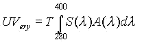
where A(lambda) is the erythemal action spectrum (CIE, 1987), S(lambda) is the spectral irradiance and T is the exposure period.
Erythemal Exposures
An upright rotating manikin was employed to model random movements of a human in an upright position. The manikin was rotated on a portable rotating base at a speed of 1 revolution per minute in an open unshaded area. The human body has a complicated topography and the range of human movements and orientations are very varied and complicated. Airey et al (1995) and Holman et al (1983) have compared UV exposures to head forms and body forms with exposures to humans and found that exposures to manikins may be related to human UV exposure. In this paper, only the standing position is employed and no other positions of the body are considered.
Polysulphone dosimeters were placed at 23 sites on the manikin, namely: vertex of the head, forehead, nose, chin, left cheek, left ear, right cheek, right ear, neck, left shoulder, right shoulder, left forearm, right forearm, lumbar spine, lower sternum, left thigh front, left thigh back, right thigh front, right thigh back, left shin front, left shin back, right shin front and right shin back at a sub-tropical latitude in Toowoomba (27.5 oS, 151.9 oE), Queensland, Australia. The erythemal exposures were measured in winter for a ten hour period from 07:00 Australian Eastern Standard Time (EST) to 17:00 EST and for a one hour period near solar noon from 11:00 EST to 12:00 EST on the 13 August 1996. The average cloud cover on this day was zero and the average solar zenith angle between 11:00 EST and 12:00 EST was 42.5�. During the course of the experiment from 07:00 EST to 17:00 EST, a human subject wore two dosimeters, one on the left shoulder, the other on the lower sternum for comparison of the exposures measured on a human to those measured on the manikin at these sites.
Body Zones’ Exposures
The human body was split into four zones, namely, head, trunk, arms and legs. Each zone of the body was divided into a 1 cm2 grid in order to employ the exposures to the sites in the previous sub-section to estimate the distribution of UV exposure. The influence of grid size has been determined by previous research (Kimlin et al 1997) with a 1 cm2 grid providing acceptable results. In a simplified model, the trunk, arm and leg zones were considered as cylinders with the appropriate radii and heights. For each exposure period, the exposures at each dosimeter site were linearly interpolated firstly in a vertical direction and secondly in a horizontal direction to provide exposures at each grid point. The head was treated in a similar manner to Kimlin et al (1997). The total exposure for each zone was calculated by summing the products of the exposures to each grid and the area of each grid. This exposure was divided by the total surface area of each zone to provide the exposures per unit area of the zones.
Body Exposure Distribution
The exposures to each grid for each of the zones were combined to provide the whole body exposure distribution. The total body exposure was calculated by summing over the whole body the product of the individual exposures to each grid and the area of each grid. The exposure was divided by the total surface area of the body area to provide exposure per unit area of the body.
Results
Erythemal Exposures
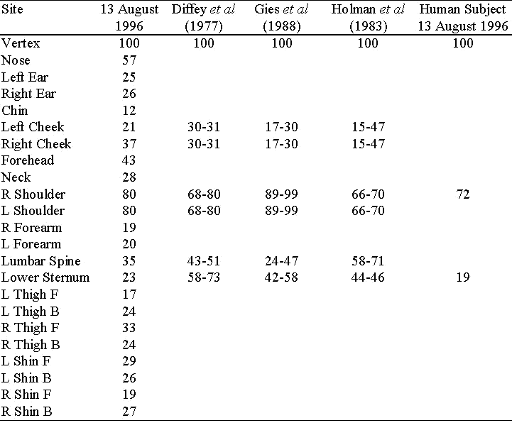
The erythemal exposures relative to the ambient exposure as measured on the vertex of the head are shown in Table 1. These are compared to those measured in other studies. The UV to the body sites from this study is within the results gained by previous investigators, except for the lower sternum site. This difference may be due to variations in the solar zenith angle which has an effect on the relative exposures to body sites. This research was undertaken at a sub-tropical latitude whereas the other studies were at high latitude locations. The exposures to the right shoulder and lower sternum of the human subject on 13 August were slightly less than those on the manikin, but well within the 20% error associated with solar erythemal measurements using polysulphone film (Diffey 1984).
Table 1 - Relative erythemal UV exposures for body sites on 13 August 1996 compared to the values obtained by previous investigators.
Body Zones’ Exposures
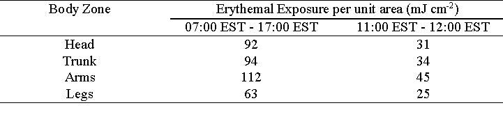
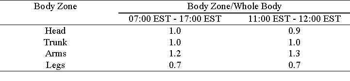
The erythemal UV exposures per unit area for each body zone from 07:00 EST to 17:00 EST and 11:00 EST to 12:00 EST on 13 August 1996 are shown in Table 2. A point to note from the table is that the arms receive an exposure about 20% - 40% more than the head or the trunk. The high exposures per unit area obtained on the arms may be due to the arms being in a position other than directly alongside the trunk. The head area receives a similar exposure per unit area to the trunk, while the legs received the lowest exposure per unit area (about 25% - 35% less than the head or the trunk).
The majority of the exposure during this day was between 11:00 EST and 12:00 EST, with the body zones in this period receiving 34% to 40% of the exposure received between 07:00 EST to 17:00 EST. All the exposures per unit area in Table 2 are over 1 minimum erythemal dose (MED), with one MED defined as 20 mJ cm-2 (Diffey 1992) and is the amount of biologically effective UV required to produce barely perceptible erythema after an interval of 8 to 24 hours following UV exposure. On the arm zone from 07:00 EST to 17:00 EST, the exposure was approximately 5.5 MED. The National Health and Medical Research Council of Australia (NHMRC 1989) has provided an occupational standard for UV exposure. The exposures to all of the body zones measured in this research for both the ten hour and one hour period are in excess of 1 MED. This is of concern as the one hour period is at approximately noon which can be associated with meal breaks when indoor workers and school children may spend a proportion of their meal breaks exposed to solar radiation and as a result exposed to high levels of UV to any unprotected body zones. Additionally, these high exposures have been measured on a winter day.
Table 2 - Erythemal UV exposures per unit area for each body zone.
Body Exposure Distribution
The exposures to each grid for each body zone have been combined to provide the whole body distribution of erythemal UV in Figure 1 on the 13 August 1996 from 07:00 EST to 17:00 EST and from 11:00 EST to 12:00 EST. The UV exposure is highest on the vertex of the head with the level of exposure decreasing for positions below the vertex of the head. The exposures per unit area to each of the body zones in Table 2 have been employed to calculate the exposure to the whole body per unit area. These are 90 mJ cm-2 for the ten hour period and 34 mJ cm-2 for the one hour period. The ratios of the exposure per unit area to each zone to the whole body exposure per unit area are provided in Table 3 for each period. In both periods, the leg zone received less per unit area than the whole body and the arm zone received more per unit area than the whole body. The possible explanation for the higher exposure to the arm zone is due to the position of the arms with respect to the other body areas whereas the legs are in a vertical orientation.
Figure 1 - Distribution of erythemal UV to the body on 13 August 1997 from 07:00 EST to 17:00 EST and 11:00 EST to 12:00 EST.
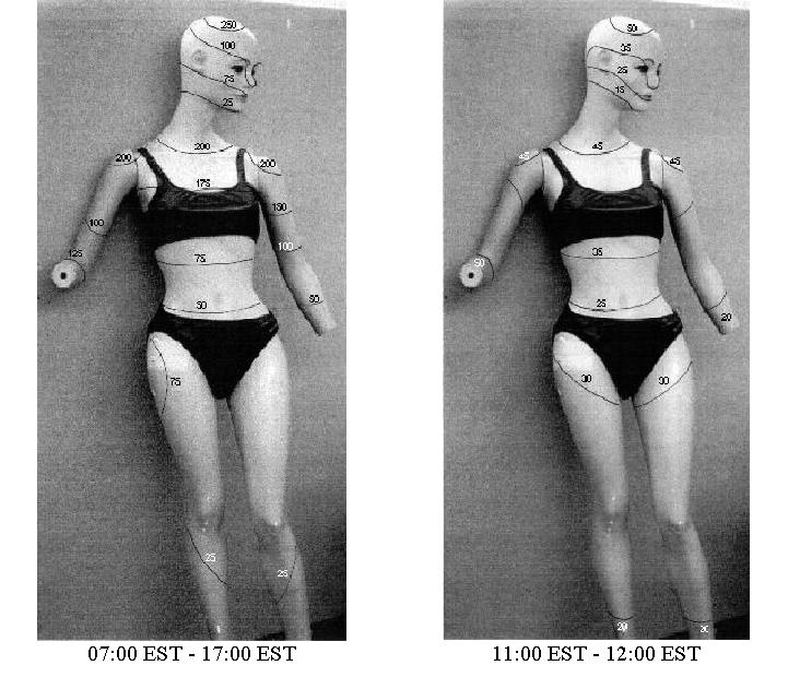
Table 3 - Ratios of the body zone exposures to the whole body exposure.
Discussion
A technique for the estimation of the erythemal UV exposure at any location on the human body using only 23 dosimeters was presented. The method presented provides the complete picture of the body distribution of erythemal UV. The distributions produced provide at a glance the parts of the body that are high skin cancer risk areas requiring appropriate protective measures. Similar exposures per unit area were received to the head zone compared to the trunk zone with the leg zone receiving 25 to 30% less compared to the head zone and the arm zone receiving 20 to 40% more compared to the head zone. The whole body exposures per unit area at the sub-tropical latitude site of this research were high at 34 and 90 mJ cm-2 for the one hour and ten hour periods respectively and highlighted the need for UV protective measures even on a winter’s day
The exposures to each of the body zones may be more suitable to be employed as cancer risk indicators as they provide the overall picture rather than the individual site exposures which may vary from site to site. Additionally, the exposures to the body zones can be employed in establishing the effectiveness of protective devices. Specifically, the exposures to the face, trunk, arm and leg zones can be employed in determining the effectiveness of hatwear, garments, sleeved clothing and shorts, pants or women’s skirts.
Acknowledgments: This project is partially supported by the Qld. Health. One of us (MK) would like to acknowledge the financial support from the Qld. Health in the form of a training scholarship.
References
Airey, D.K., Wong, J.C.F. & Fleming, R.A. 1995, “A comparison of human- and headform- based measurements of solar ultraviolet B dose,” Photodermatol. Photoimmunol. Photomed. vol.11, no.4, pp.155-158.
CIE (International Commission on Illumination) Research Note 1987, A reference action spectrum for ultraviolet induced erythema in human skin, CIE J. vol.6, pp.17-22.
Diffey, B.L., Kerwin, M. & Davis, A. 1977, “The anatomical distribution of sunlight,” Br. J. Dermatol., vol.97, pp.407-410.
Diffey, B.L. 1984, “Personal ultraviolet radiation dosimetry with polysulphone film badges,” Photodermatol., vol.1, pp.151-157.
Diffey, B.L. 1989, “Ultraviolet radiation dosimetry with polysulphone film”, in Radiation Measurement in Photobiology, ed. B.L. Diffey, pp.136-159, Academic Press, New York.
Diffey, B.L. 1992, “Stratospheric ozone depletion and the risk of non-melanoma skin cancer in a British population,” Phys. Med. Biol., vol.37, no.12, pp.2267-2279.
Diffey, B.L. & Cheeseman, J. 1992, “Sun protection with hats,” Br. J. Dermatol., vol.127, pp.10-12.
Gies, P., Roy, C. & Elliot, G. 1988, “The anatomical distribution of solar UVR with emphasis on the eye,” Proc. 7th Congress of the International Radiation Protection Association, vol.1, 10-17th April, pp.341-344.
Gies, P., Roy, C., Toomey, S., MacLennan, R. & Watson, M. 1995, “Solar UVR exposures of three groups of outdoor workers on the Sunshine Coast, Queensland,” Photochem. Photobiol., vol.62, no.6, pp.1015-1021.
Herlihy, E., Gies, P.H., Roy, C.R. & Jones, M. 1994, “Personal dosimetry of solar UV radiation for different outdoor activities,” Photochem. Photobiol., vol.60, no.3, pp.288-294.
Holman, C.D.J., Gibson, I.M., Stephenson, M. & Armstrong, B.K. 1983, “Ultraviolet irradiation of human body sites in relation to occupation and outdoor activity: field studies using personal UVR dosimeters,” Clin. Exp. Dermatol., vol.8, pp.269-277.
Kimlin, M.G., Parisi, A.V. & Wong, J.C.F. 1997, “The facial distribution of erythemal ultraviolet exposure in south east queensland,” in press Phys. Med. Biol.
Longstreth, J.D, de Gruijl, F.R., Kripke, M.L., Takizawa, Y. & van der Leun, J.C. 1995, “Effects of increased solar ultraviolet radiation on human health,” Ambio, vol.24, pp.153-165.
McCarthy, W.H. & Shaw, H.M. 1989, “Skin Cancer in Australia,” Med. J. Aust., vol.150, pp.469-470.
National Health and Medical Research Council, 1989, “Occupational standard for exposure to ultraviolet radiation,” Radiation Health Series No.29. NHMRC, Canberra.
Rosenthal, F.S., West, S.K., Munoz, B., Emmett, E.A., Strickland, P.T. & Taylor, H.R. 1991, “Ocular and facial skin exposure to ultraviolet radiation in sunlight: a Personal exposure model with application to a worker population,” Health Phys., vol.61, pp.77-86.
Wong, C.F., Fleming, R.A., Carter, S.J., Ring, I.T. & Vishvakarman, D. 1992, “Measurement of human exposure to ultraviolet-B solar radiation using a CR-39 dosimeter,” Health Phys., vol.63, no.4, pp.457-461.
Wong, C.F., Toomey, S., Fleming, R.A. & Thomas, B.W. 1995, “UV-B radiometry and dosimetry for solar measurements,” Health Phys., vol.68, no.2, pp.175-184.
Wong, C.F., Airey, D.K. & Fleming, R. 1996, “Annual reduction of solar UV exposure to the facial area of outdoor workers in Southeast Queensland by wearing a hat,” Photodermatol. Photoimmunol. Photomed., vol.12, pp.131-135.