MATERIALS AND METHODS
Linoleic acid (95%), methyl linoleate (95%), tris[hydroxymethyl]aminomethane (TRIS) buffer, polyoxyethylene 9 lauryl ether (lubrol-PX), and D-glucose were obtained from Sigma Chemical Company; FeCl2.4H2O and CuCl2.2H2O were purchased from BDH Chemicals Ltd; and nitrilotriacetic acid from Aldrich Chemical Company. Oxygen used during these experiments was medical grade (99.5% O2) and nitrogen used was instrument grade (99.99% N2). Distilled water was used in all solutions.
Preparation of Assay Buffer: Initially a 0.1 M TRIS solution was prepared in water. The pH of this buffer was adjusted to 8 with 0.1 M hydrochloric acid. A dispersant, lubrol-PX, was added to the TRIS/HCl pH 8 buffer at a concentration of 0.8%. This TRIS/HCl/lubrol-PX buffer was determined to be optically clear in the 210-800 nm wavelength range.
Chemiluminescence measurement
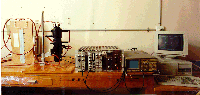
The photon counting equipment used to measure the photon emission from linoleic acid (LA) or methyl linoleate (ML) samples (Figure 1) has been described in more detail elsewhere (21,22), and utilized a 13 dynode EMI 9635 QA photomultiplier (PM) tube sensitive in the wavelength range 200-630 nm. The PM tube, its dynode chain and a Pyrex sample cuvette were housed in a light-tight thermostatted (25.0 ± 0.1°C) container. The end window of the PM tube was situated 8 mm directly below the sample cuvette and could be isolated by means of a shutter. Power to the dynode chain was supplied by an Ortec model 556 stabilized high voltage power supply and the photoelectron pulses were amplified and discriminated from noise pulses by an Ortec 716A amplifier and an Ortec model 406A single channel analyzer, respectively. Output from the single channel analyzer was collected by and processed on a microcomputer (using PC 3-Channel Counter, Fozdar Computing).
Figure 1
Light detection system
enlarged view
For the experiments carried out at 60°C approximately 0.5 cm3 of either LA and ML was placed in a brown glass sample vial, which was held in a water bath kept at a constant 60°C. The samples were kept under an oxygen atmosphere for the duration of each experiment. Initially, and then every 24 h, an accurately weighed portion of the sample was taken from the vial and dissolved with buffer to form a 2 mg/cm3 optically clear micellar solution. 20 cm3 of this solution was then injected into the sample cuvette of the photon counter. The mean count rate above background was determined whilst oxygen was bubbled through the solution at 0.5 cm3 s-1. For experiments which involved either glucose or iron (II)-nitrilotriacetate, a 100 cm3 solution of fresh 2 mg/cm3 LA or ML was prepared with either 20 mM glucose or 200 mM Fe2+-NTA. This solution was then injected into the cuvette of the photon counter. Oxygen was bubbled through the solution at 0.5 cm3 s-1 and CL was averaged over each minute and then recorded for the duration of each experiment.
Lipid Peroxidation Assay
The peroxidation of lipids can be measured as an increase in absorbance of ultra-violet light due to conjugated diene formation. The concentration of dienes can hence be calculated using an extinction coefficient. The amount of conjugated dienes is directly proportional to the amount of hydroperoxides formed as the major initial product of the lipid peroxidation reaction. The hydroperoxides eventually break down to form the final products, so this method is a measure of the initial stages of the reaction. For linoleic acid and methyl linoleate the peak absorption wavelength is 234 nm and the extinction coefficient is 28 x 106 cm2 mol-1. For this study, UV absorption spectroscopy was chosen as the method to measure lipid peroxidation. It was chosen for its simplicity of measurement and its specificity. A direct measure of the concentration of conjugated dienes is obtained by this method, which in the presence of oxygen is directly related to the amount of hydroperoxides formed.
Lipid peroxidation was thus monitored by measuring conjugated diene formation as absorption of UV light at 234 nm. The absorbance of light at 270-280 nm was also monitored. This absorbance may be due to secondary oxidation products such as aldehydes and ketones, and hence may be an indication of the formation of the final products of lipid peroxidation. At certain times during the experiments, solutions of 0.04 mg/cm3 LA or ML were prepared by taking a sample of the oil at 60oC or by withdrawing 1 cm3 of the solution from the cuvette in the photon counter and diluting appropriately with buffer. UV absorption measurements were carried out on a Hewlett Packard 8452 Diode Array Spectrophotometer.
Sample Quality: Throughout all the 60°C, glucose, and Fe2+-NTA experiments, the samples were monitored for any change in smell or colour, since these changes are associated with rancidity.
 Results |
|||
 Abstract |
Introduction |
 Discussion |
 References |
![]()